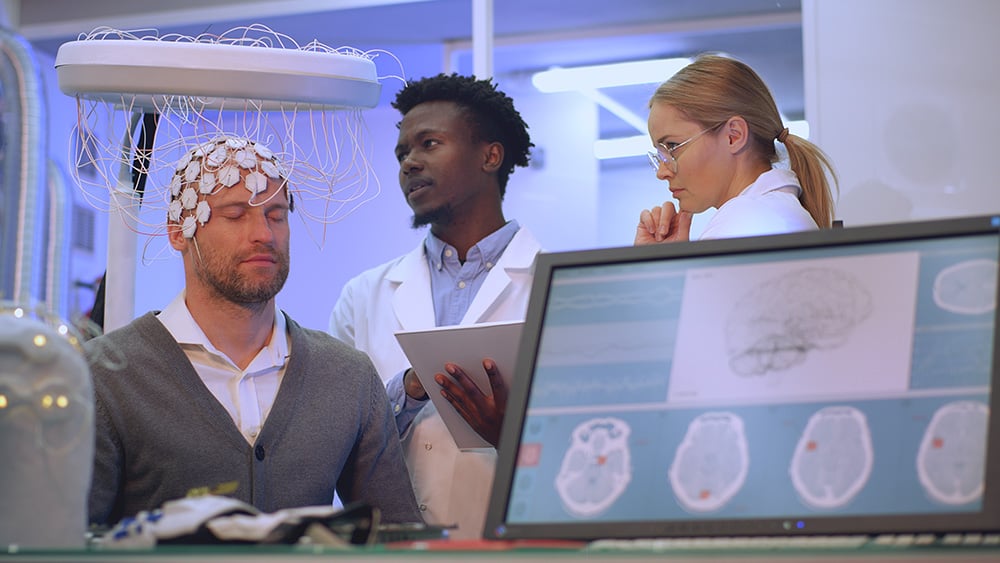
Rochester Regional Neurology offers a variety of neurology testing and diagnosis for both inpatient and outpatient services. The information obtained from neurology diagnosis testing depends on the quality of the study and on the experience and knowledge of the interpreting physician. Neurology diagnostic tests are performed with state-of-the-art equipment and test results are then analyzed by neurologists with either fellowship or advance specialty training in the interpretation of these tests. The results of your tests will be delivered promptly to your healthcare provider.
We offer the following neurology diagnostic tests:
An electroencephalogram (EEG) is a test to detect abnormalities related to electrical activity of the brain. When the brain cells send messages to each other, they produce tiny electrical signals. Your brain cells communicate via electrical impulses and are active all the time, even when you’re asleep. This activity shows up as wavy lines on an EEG recording. An EEG is one of the main diagnostic tests for epilepsy. An EEG may also play a role in neurology diagnosis other disorders.
In an EEG test, electrodes are placed onto your scalp using a sticky substance. These electrodes pick up the electrical signals from your brain and send them to an EEG machine, which will record the signals as wavy lines onto a computer. The EEG machine records the brain’s electrical activity as a series of traces. Each trace corresponds to a different region of the brain. Photic Stimulation and Hyperventilation are two procedures usually done as a part of routine EEG test and can provoke seizure in certain percentage of patients. This neurology test is a painless procedure that takes anywhere from an hour to an hour and a half to complete.
Your carotid arteries carry blood and oxygen to your brain. A carotid ultrasound takes pictures of your carotid arteries. This test is to check your arteries for blockage or narrowing due to the buildup of plaque or cholesterol. If a blockage or narrowing is present, then your risk of having a stroke may be higher.
Warm ultrasound gel will be placed on each side of your neck. The sonographer will move a small microphone over the gel to take pictures of your arteries while listening to the blood flow. This neurology diagnosis test takes anywhere from a half hour to an hour to complete.
Transcranial Doppler (TCD) is a test that uses sound waves to study the blood flow in the arteries in your brain. As the blood flows through these arteries it makes sounds that the sonographer listens to and records. This test may help to determine possible causes of stroke, for example, an abnormal connection between your arteries and veins.
Warm ultrasound gel will be placed on your closed eyelids, on a small area next to each of your ears, and then on a small area on the back of your head. The sonographer will listen to the arteries behind those areas with a small microphone. This neurology testing takes anywhere from a half hour to an hour to complete.
Additional TCD studies can also be used to identify specialized problems related to brain blood flow:
During this test, a cap is placed on the head which has a microphone attached to each side. The microphone focuses on two arteries in the brain. Your sonographer will listen to the blood flowing through these arteries and record the results. The test will take 30-45 minutes to complete.
During this test, a cap with microphones on each side is placed on your head. A temporary IV is placed into a blood vessel in your arm and a salt solution containing tiny bubbles is injected. The sonographer will listen to the blood flow through the arteries in your brain to detect if any of the bubbles reach the brain. The test takes 30 to 45 minutes to complete.